ZEISS Xradia CrystalCT

The first commercially available crystallographic imaging microCT system.

ZEISS Xradia CrystalCT is your ground-breaking microCT for unlocking the crystallographic and microstructural secrets of your samples.

It uniquely augments the powerful technique of computed tomography with the ability to reveal crystallographic grain microstructures, transforming the way polycrystalline materials (such as metals, additive manufacturing, ceramics, etc.) can be studied, leading to newer and deeper insights into materials research.

Perform non-destructive mapping of grain morphology in 3D.
Characterize materials such as metals, alloys, and ceramics.
Map larger volumes and a wider array of sample geometries at higher throughput.
Advance materials characterization and discovery through ground-breaking diffraction scanning modes.
Achieve superior sample representivity to create high fidelity computational models.

Technology Insights
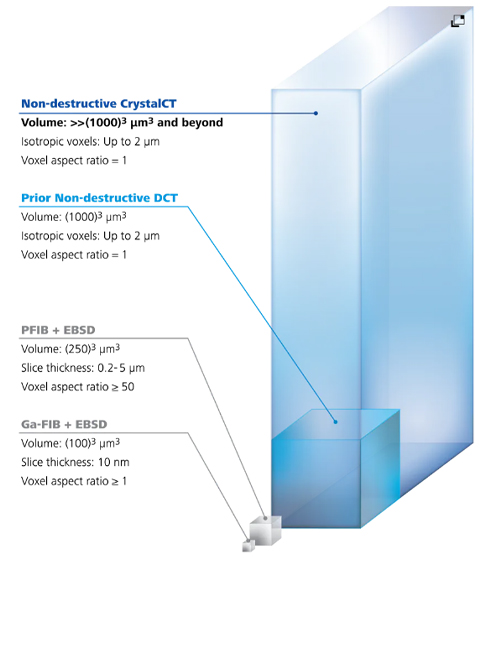
Super Sample Representivity

Sample representivity – obtaining large volumes of real data to create high fidelity computational models – has been a challenge for crystallographic imaging.
ZEISS Xradia CrystalCT offers advanced DCT modes that overcome some of the previous challenges of conventional DCT data collection that assumes the ROI in the sample is fully illuminated by the aperture field of view (FOV) for all rotational angles of the sample.

ZEISS Xradia CrystalCT advanced diffraction scanning modes include

✓ Helical Phyllotaxis
Helical phyllotaxis rotation is used for long aspect ratio cylindrical samples.
✓ Helical Phyllotaxis Raster
Helical phyllotaxis raster is used for samples that are typically wider than the field of view.
Helical Phyllotaxis HART
Phyllotaxis with high aspect ratio tomography, or HART, solves the problem of flat or plate-like sample imaging.

Technical Specifications